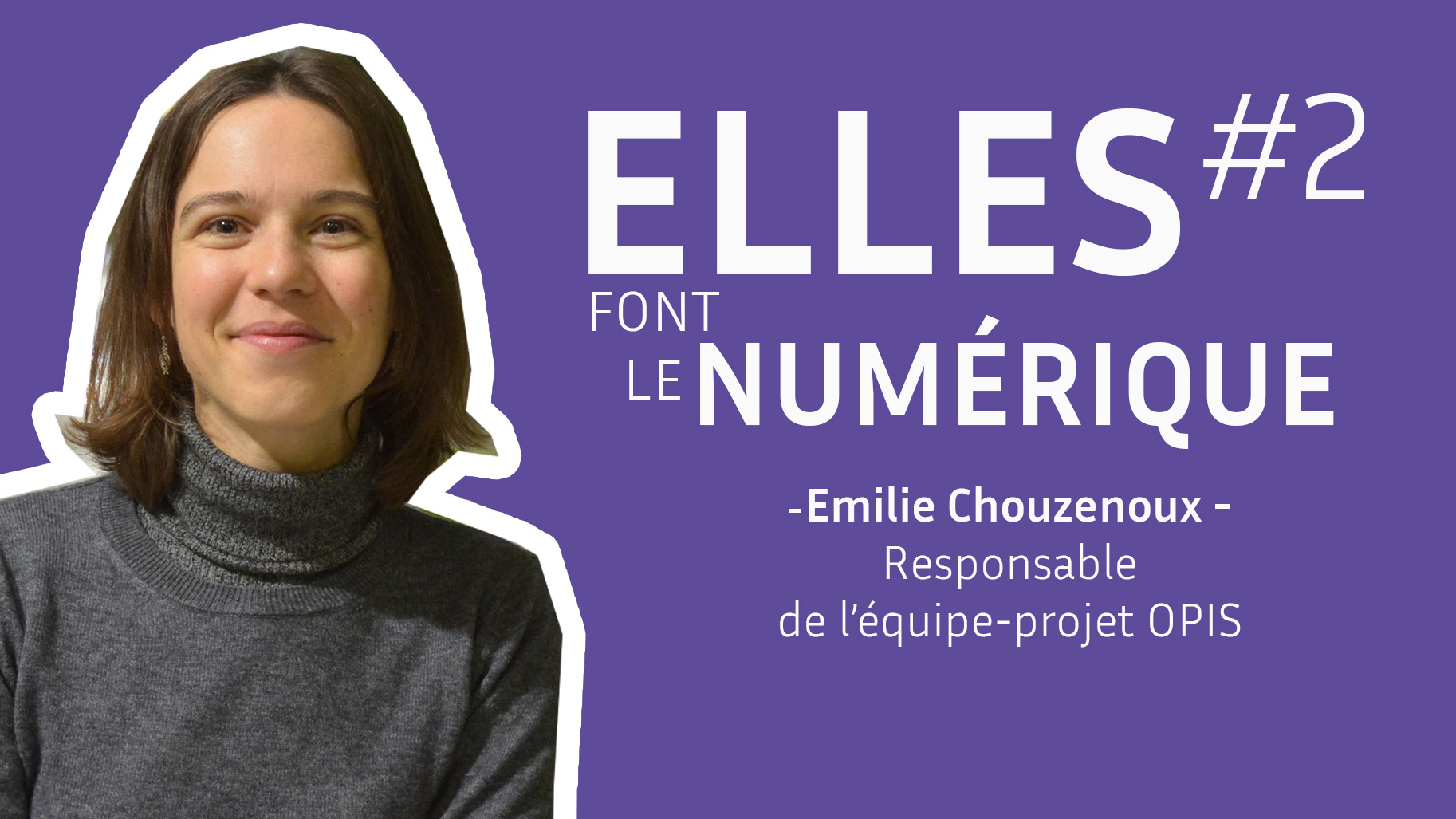
From algorithms to diagnosis: the exceptional career of Émilie Chouzenoux
Date:
Changed on 04/04/2025

I trained as an engineer in automatic control and signal processing at the École Centrale de Nantes. Like most students, it took me a few years to decide on the career and specialism I wanted to pursue. I have always been attracted to mathematics and its applications in the medical field, and had the desire to teach in order to disseminate knowledge.
I therefore explored several different avenues. Through my internships in clinics and laboratories, I met researchers and engineers and discovered a number of specialisms applied to the medical field. It all fell into place in my final year when I discovered the field of medical imaging research and the problems associated with image reconstruction, which fascinated me from the outset. This topic combines mathematical developments and algorithmic implementations with concrete applications enabling the direct visualisation of results.
It was during my thesis that I perfected my expertise in mathematical optimisation, which is now central to my research and scientific contributions. My career path then led me to a position as a lecturer at Paris-Est University, before I joined Inria in 2016 as an associate researcher, becoming a researcher in 2019. In 2023, I was promoted to Senior Researcher at Inria, and took charge of the OPIS project team, which was a major step forward in my career. Leading a team is an exciting challenge that has taught me a lot about management, organisation, interpersonal skills and communication. It complements my skills as a researcher perfectly.
The researchers in the Optimisation, Imaging and Health (OPIS) project team are studying three topics:
This team is shared by three entities: the Inria Saclay Centre, CentraleSupélec and Paris-Saclay University at the CVN laboratory (Digital Vision Centre).
The team is collaborating with a number of hospital groups in the Ile-de-France region: AP-HP, Gustave Roussy Hospital, Saint-Joseph Hospital; and a number of manufacturers, including GE Healthcare and Essilor.
I see two main challenges facing women scientists.
The first is self-censorship. I have sometimes hesitated – and that is still the case – to apply for prizes or competitions, thinking that I don't have the ideal profile. Thankfully, I have been fortunate enough to be surrounded by caring colleagues who have encouraged me to apply and to believe in my abilities. Having a good support network is essential for breaking down the barriers that may sometimes be self-imposed.
The other challenge relates to managing a predominantly male team. As a female manager, I have noticed that my PhD students sometimes confide in me more than my male colleagues with regard to personal issues. It's important to remind them of the professional context and that, although I'm a good listener, my main role is to act as their PhD supervisor and team leader. That said, in my day-to-day work, I see greater diversity in my collaborations with the medical profession, where I work with numerous female heads of departments and radiologists
I develop medical-image-reconstruction algorithms. When a scanner acquires data, it does not directly produce a usable image, but rather noisy sensor data that is impossible to interpret. An image of the highest possible quality must be reconstructed as quickly as possible, while ensuring that it is an accurate reflection of the clinical reality. The aim is to use this raw data to produce accurate images that can be interpreted by doctors to guide diagnoses and decision-making. And all without any delay. Patients in the radiologist's waiting room cannot hang around for hours. We even work in interventional imaging contexts, where the image must be displayed immediately for direct interpretation by the surgeon. This requires the estimation of millions of variables and therefore the resolution of a mathematical optimisation problem on a massive scale.
One of our missions is to provide theoretical analyses – a form of certification – to explain and improve the understanding of what can be done in clinical practice. For example, I have worked with GE Healthcare on a study examining a key stage in the reconstruction of scanner images: retro-projection. We mathematically characterised the consequences of approximating this stage, and provided rules to ensure the reliability of the final images. Three publications have had a major impact in my field: "Solution of Mismatched Monotone+Lipschitz Inclusion Problems", "Convergence Results for Primal-Dual Algorithms in the Presence of Adjoint Mismatch", "Unmatched Preconditioning of the Proximal Gradient Algorithm". The findings have had a direct impact on the quality of diagnoses and healthcare professionals’ confidence in these technologies.
This award recognizes Emilie Chouzenoux's outstanding contributions to the field of signal and image processing optimization, with a successful application to medical imaging. “This prize is very important to me. It's recognition of the work I've accomplished over the years. It validates, through my peers, the impact and visibility of my research on a European scale. I'm also proud that this prize has been awarded to a woman. We're still too much in the minority at these scientific conferences, and it shows the importance of networking and solidarity between women scientists. This kind of recognition encourages other young female researchers to persevere and believe in their skills.”
One of my recent projects, in collaboration with St Joseph Hospital and Saint-Antoine Hospital at AP-HP (the Paris University Hospital Trust), sets out to use AI to provide guidance for the diagnosis of intestinal obstructions in patients admitted to emergency departments. Just imagine, it's 3 a.m. in the emergency department. A patient has been admitted with extremely severe pain and the radiologist has to scan the entire body and make a decision about surgery with major consequences for the patient's life. Image analysis is a time-consuming and very difficult process. Our initial results show that our AI model can detect an occlusion as effectively as an expert radiologist.
We are now working with our radiology partners on the crucial question of the need for surgery (this type of surgery can lead to serious complications), so as to help practitioners make faster, more accurate decisions.
I am increasingly interested in the hybridisation of traditional mathematical optimisation and artificial intelligence. The challenge is to go beyond simply displaying the image and move increasingly towards providing diagnostic assistance.
I would like to develop AI tools that can be used at the patient’s bedside, but we still have a long way to go. The clinical acceptability of AI remains a major obstacle. There is considerable mistrust of these methods, not least because of their lack of transparency and interpretability. To overcome these obstacles, we need to succeed in creating algorithms capable of explaining their decisions and providing confidence intervals (a meaningful percentage of certainty about the result), to enable healthcare professionals to use them with confidence.