Research
Using Statistics to Pierce the Mysteries of the Brain
Tractography showing the connections between the different regions of the brain
-
© Inria / Photo C. Morel
Nearly 100,000 billion neurons, each typically having 10,000 synapses and an almost infinite number of possible connections: grey matter is, in itself, a formidable mathematical object. At theInstitut du cerveau et de la moelle épinière(the Brain and Spinal Cord Institute),the researchers of the Aramis project team, using statistics and image analysis, work with doctors to improve our understanding of the mechanisms of pathologies such as Alzheimer’s disease.
At the heart of the Pitié-Salpêtrière Hospital in Paris, among century-old brick buildings, stands a vessel of glass. Located since 2010 in the midst of the largest hospital complex in the French capital,the Institut du Cerveau et de la Moelle épinière(ICM) houses more than 600 researchers from all over the world. Here, neurobiologists rub shoulders with specialists in the cognitive sciences, mathematicians, statisticians and computer scientists.
Computer scientists at the hospital
An Inria-CNRS-Inserm-Université Pierre et Marie Curie joint project team that goes by the name of Aramis has taken up residence on the 3rd floor of this 22,000 sqm building. In a hi-tech environment, equations and mathematical formulae inscribed in felt-tip decorate the glass walls of the offices. Four researchers and thirty or so PhD students, post-docs and engineers work here day in day out alongside doctors, neurologists and neuroradiologists. They share a common goal: to study and understand the structure and functions of the human brain. They are studying the network of neurons and fibres that connect the different regions of the brain to each other as a very complex geometrical form. “Methods of statistical analysis help us to measure the physiological consequences of certain neurodegenerative diseases”, explains Olivier Colliot, the project team leader, indicating the MRI images displayed on his computer. One image shows a patient suffering from Alzheimer’s disease, while another MRI scan is from a healthy patient. In the first, the hippocampus is smaller. “This atrophy is a biomarker of the disease, and it can be used to monitor how patients evolve. Because the loss of volume is between 3 to 5 % per year, the progress of the atrophy over time is difficult to appreciate with the naked eye. These changes can only be detected through very precise measurements, using special methods of analysis. Perhaps one day they will enable us to provide earlier diagnoses.” The Aramis research team works closely with doctors to help form a clearer understanding of diseases that remain mysterious. This is so with frontotemporal dementia, a rare condition that can take very different forms in different patients. Some develop behavioural disorders; others suffer from disorders of language or memory. “We use mathematics to try to analyse how the same biological cause – in this case a genetic mutation – can produce symptoms that are so different”, explains Olivier Colliot.
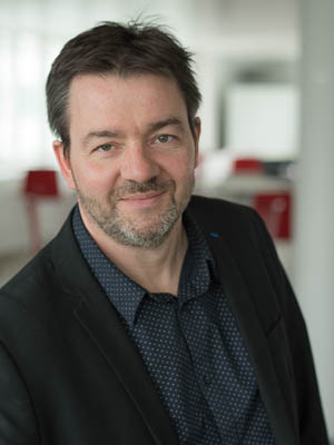
Fabrizio De Vico Fallani, Olivier Colliot (Dir.) and Stanley Durrleman, Aramis team researchers – Inria, Université Pierre et Marie Curie, INSERM and CNRS joint research team, within the ICM – Institut du Cerveau et de la Moelle épinière.
-
© Inria / Photo C. Morel
Statistical methods model the evolution of neurological diseases
With the aging of the population, Alzheimer’s disease, rare before age 65, is becoming a public health challenge. Stanley Durrleman has chosen to direct his research to this pathology in particular. He has received funding from the European Research Council for a 5-year period set to start in September 2016. “Studying this disease requires a change of paradigm”, explains the researcher. “Normally, when we talk about illness, either we are affected by it or we’re not. With Alzheimer’s, it is more complicated, the disease is superimposed on aging. And of course, it can start many years before the appearance of the first symptoms.” In the face of such a complex time span, Stanley Durrleman has chosen to design methods of statistical analysis based on longitudinal images. Rather than compare a group of healthy patients with a group of sick patients, he studies the changes in cerebral structure and function over time in the same patients. His aim is to typify the change in brains affected by the disease and identify a set of possible variations. Alzheimer’s disease does not develop at the same rhythm in all affected patients: The condition may appear earlier or later, and develop more or less quickly.”To make sense of patient data, we need to find the right criteria, because age doesn’t work. We are going to have to find a way to standardise the aging trajectories that are specific to each patient”, he explains. “It is a real challenge, because for the moment, the methodological framework does not exist. We have to invent it all!”
Graph theory at work to improve our understanding of the brain
Researcher Fabrizio De Vico Fallani studies the brain as a network: The nodes correspond to the cortical regions, i.e. to areas of the brain that communicate with each other. The brain thus becomes a mathematical object to which graph theory can be applied. “The brain contains billions of neurons and we do not yet have the technical resources to reproduce this network in its totality. But we are capable of distinguishing the major regions and of observing how they interact.” The researcher has also helped to develop a brain-machine interface. The goal is to use the power of thought alone to point the cursor towards a target, but the software cannot be used properly without practice. “If we just think about moving our hand when it is at rest, the brain transmits an electrical signal similar to that transmitted when the body actually moves. The same thing happens with people who are paralysed: they have lost their mobility, but their brain still functions.” We can exploit these electrical signals, but using this type of interface does not come naturally. “So we are studying how the brain adapts and how it can learn to use these tools.” The young researcher has also received funding for his work from the FrenchAgence Nationale de la Rechercheand the National Science Foundation.
A tool that lets grey matter speak
Based on his theoretical results, Fabrizio De Vico Fallani is also developing tools capable of exploiting certain unexplored capacities of the brain. These tools may enable people who are paralysed or deprived of the power of speech to communicate with the outside world. The system that he has designed is truly futuristic. Imagine yourself in front of a television screen, wearing a cap covered with electrodes. Each of them is connected to a machine that analyses the electrical signals transmitted inside your skull. On the screen, the 26 letters of the alphabet are organised in lines and columns. Fabrizio De Vico Fallani asks you to think of a word and then runs the system. A light bar first highlights each of the columns one by one, and then the lines. You have nothing to do except think of the first letter of your word while watching the screen closely. When the bar highlights the column containing the letter concerned, the brain transmits an electrical signal without your being able to control it. It does the same thing when the line containing the letter is highlighted; the system can then deduce the letter you are thinking about. The system allows all the possible words to be spelled out quite quickly. “I am doing research in applied mathematics, so what I’m interested in essentially is developing theoretical models of the operation of the brain. But knowing that our work could one day be useful to disabled people is extremely gratifying.”
Reportage dans l’équipe ARAMIS, équipe pluridisciplinaire regroupant des chercheurs spécialistes des sciences informatiques, de traitement du signal, et des experts médicaux en neurologie et en imagerie médicale.
ARAMIS est une équipe de recherche commune à Inria, l’Université Pierre et Marie Curie, INSERM et le CNRS, au sein de l’ICM – Institut du Cerveau et de la Moelle épinière.
© Inria / Photo C. Morel
Keywords: Inria de Paris Maladies Neurologie Pitié-Salpêtrière Aramis Statistiques Cerveau Graphes Modélisation